

Baseline HD IVUS measurements of proximal RCA lesion – MLA: 2.64 mm2
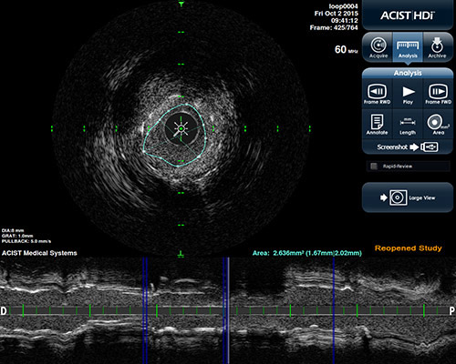
HD IVUS measurements of mid-RCA lesion – MLA: 2.85 mm2
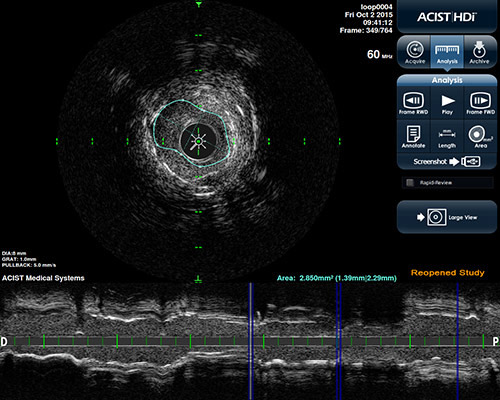
Pre-PCI HD IVUS (1) measurements of proximal lesion after 1st cutting balloon dilation – MLA: 3.54 mm2
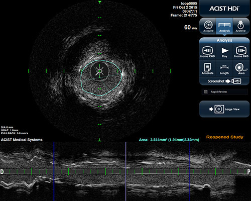
Pre-PCI HD IVUS (1) measurements of mid-lesion after 1st cutting balloon dilation – MLA: 4.026 mm2
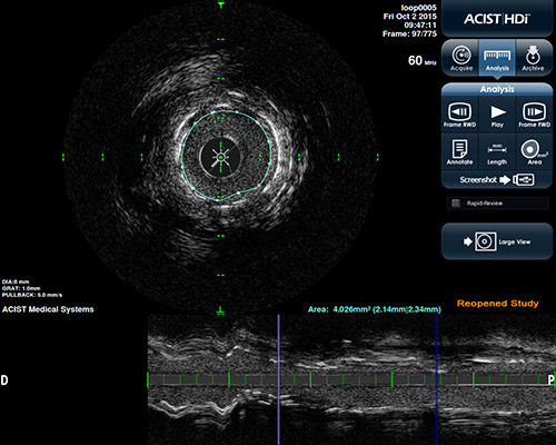
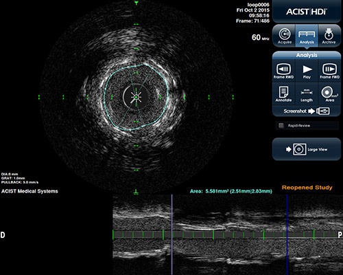
Pre-PCI HD IVUS (1) measurements of proximal lesion after 2nd NC Emerge™ PTCA Dilation Catheter2 dilation – MLA: 7.18 mm2
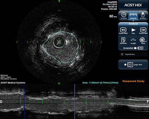
Pre-PCI HD IVUS (1) measurements of mid-lesion after 2nd NC Emerge™ PTCA Dilation Catheter2 dilation – MLA: 5.58 mm2
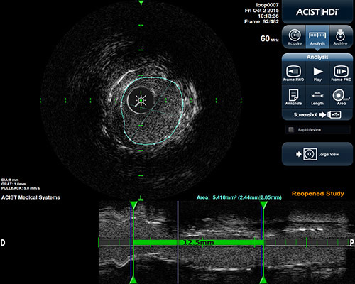
Pre-PCI HD IVUS (2) measurements of proximal lesion after laser atherectomy and NC Emerge™ PTCA Dilation Catheter dilation – MLA: 7.71 mm2
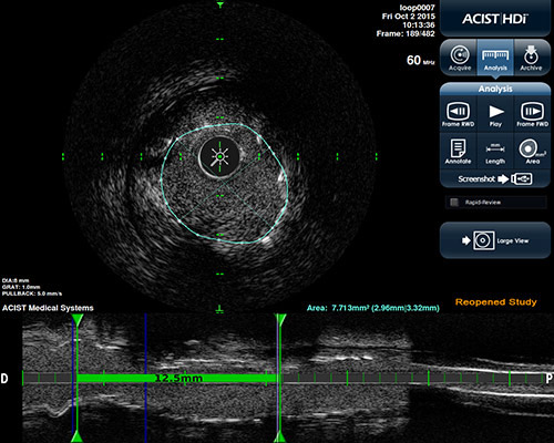
Pre-PCI HD IVUS (2) measurements of mid-lesion after laser atherectomy and NC Emerge™ PTCA Dilation Catheter dilation –MLA: 5.41 mm2
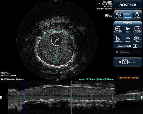
Post-PCI HD IVUS measurements of proximal lesion - MLA: 10.15 mm2
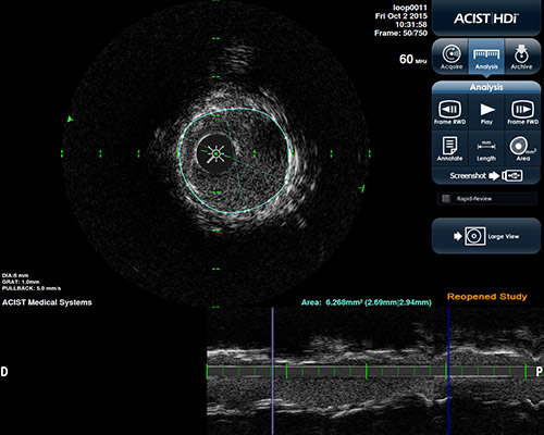
Post-PCI HD IVUS measurements of mid-lesion - MLA: 6.27 mm2
 Baseline angiography of RCA
  Baseline HD IVUS of RCA
  Pre-PCI angiography after laser atherectomy and NC Emerge™ PTCA Dilation Catheter balloon dilation
  Post-PCI angiography after post-PCI, post-balloon dilation – View 1
  Post-PCI angiography after post-PCI, post-balloon dilation – View 2
  Post-PCI HD IVUS