

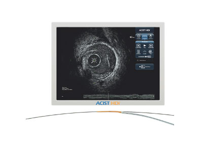
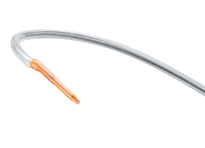
ACIST HDi High-Definition IVUS System enables optimized visualization to inform and illuminate coronary and peripheral intervention strategy through advanced imaging modes, improved deliverability of the Kodama IVUS Catheter and interactive compact console.
Click here for the HubVue™ Imaging Workflow System and PACS integration
Now indicated for both coronary and peripheral vascular procedures.
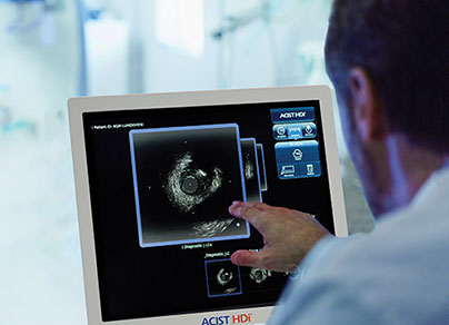
Providing you the power to see with certainty.
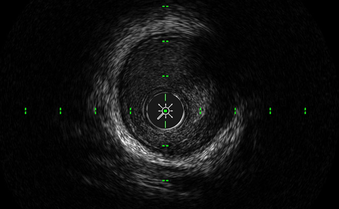
Darkens the coronary lumen for better border detection.
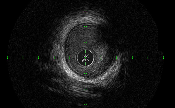
Increases gray scale for finer blood speckle, tissue and plaque differentiation.
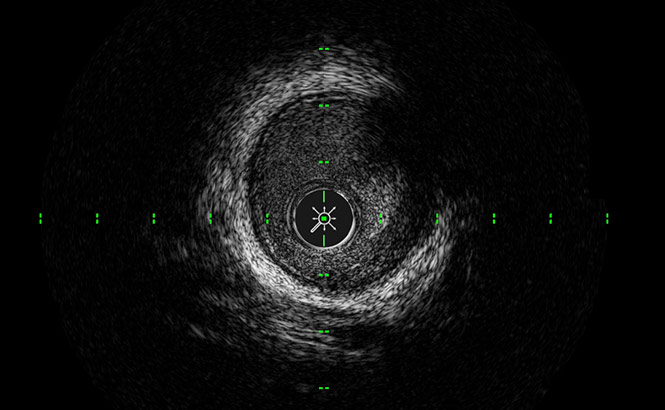
Optimizes the balance of high resolution and depth of penetration and enables full vessel wall visualization.

Better visualization of media than OCT for optimizing stent sizing4
Enables enhanced imaging by providing sufficient penetration at 60 MHz to see the media layer, even in larger plaque volumes4, so the physicians can maximize the stenting cross sectional area and may lead to better patient outcomes5
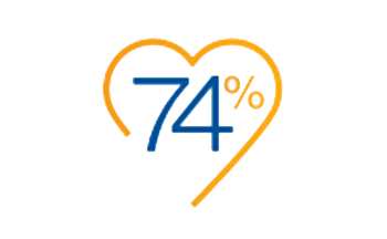
IVUS Use changed the treatment strategy during the procedure 74% of the time6
IVUS-guided stents also had low proximal & distal plaque burden, & higher MSA
compared to the angio-guided group7

8x more detection of lipid pools at 60 MHz than 40 MHz8
IVUS-guided PCI is beneficial for preventing the Stent Edge Restenosis in cases with diffused plaque lesions compared to angiography alone9

IVUS is less expensive and more cost effective than
angiography in 71% of PCI procedures10
IVUS provides improved outcomes at lower costs, with greater economic benefit especially in higher risk subgroups (diabetes, renal insufficiency, ACS)
This is based on a lifetime economic model from Italian healthcare payer perspective10
Use of IVUS in lower extremity procedures was associated with lower risk of complications post-procedure and lower risk of amputation11
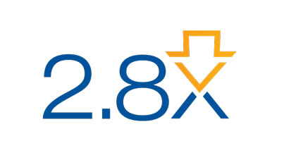
2.8x less target lesion revascularizations at one-year follow up when using IVUS in directional atherectomy procedures compared to angiography alone12

Visual estimation measurements obtained from angiographic images were consistently smaller across all arterial segments than those obtained from IVUS assessment13

69% more calcium detected by IVUS compared to angiography in the same lesions14

2x more thrombus identified by IVUS compared to angiography15

4x more post-stent dissections were identified by IVUS compared to angiography16
3.5x more post-angioplasty dissections were identified by IVUS compared to angiography17
6x more post-atherectomy dissections were identified by IVUS compared to angiography17
Rx Only
Prior to use, reference Instructions for Use, inside the product carton (when available) or at < enterwebsite.com> for more detailed information on safe use of the device.
Indications for Use:
The ACIST HDi® System is intended to be used for the ultrasound examination of coronary and peripheral intravascular pathology. Intravascular ultrasound imaging is indicated in patients who are candidates for transluminal interventional procedures. The ACIST Kodama Intravascular Ultrasound Catheter is intended for use with the ACIST HDi System.
Contraindications:
Contraindicated for patients with: bacteremia or sepsis; arterial spasm; major coagulation system abnormalities; mechanical heart valves that would be crossed by the catheter; severe hemodynamic instability or shock; total vessel occlusion (prior to initial stages of revascularization). Contraindicated for use in the cerebrovascular arteries. In coronary procedures, the product is also contraindicated for patients who are: disqualified for revascularization surgery; disqualified for balloon angioplasty (PTCA).
Important Safety Info:
Intravascular ultrasound studies using this product should be performed only by physicians and other medical professionals fully trained in the required techniques and procedures.The Kodama catheter contains a short monorail guidewire engagement system. As such, it is susceptible to guidewire entanglement and/or prolapse during catheter deployment and withdrawal. Before use and when possible during use, inspect the Kodama catheter carefully for kinks or any other damage. Do not use a kinked or damaged catheter because vessel damage and/or the inability to advance or withdraw the catheter may occur.
Never advance or withdraw the Kodama catheter against resistance until the cause of the resistance is determined by fluoroscopy. Movement of the catheter or guidewire against resistance may result in elongation or separation of the catheter or guidewire tip, damage to the catheter, or vessel perforation.
When advancing the Kodama catheter through a stented vessel, short monorail catheter designs are susceptible to guidewire/ catheter entrapment, catheter tip separation, and/or stent dislocation.
Adverse events that may occur as a consequence of intravascular ultrasound imaging include (but are not limited to): vessel occlusion and/or abrupt closure; air embolism; vessel dissection, injury, or perforation; vessel rupture, injury, or perforation; acute myocardial infarction; cardiac arrhythmias including but not limited to ventricular tachycardia, ventricular fibrillation, and complete heart block; cardiac tamponade; catheter/guidewire entrapment; catheter induced ischemia; death; vessel trauma requiring treatment/surgical intervention, including angioplasty/stent; infection; stent strut damage; stroke (including cerebral vascular accident and transient ischemic attack); thrombus formation or thromboembolism; vasospasm.
You are accessing scientific and educational content provided by ACIST Medical Systems that is intended for healthcare professionals only.
By proceeding, you confirm that you are a healthcare professional and acknowledge that you have read and accepted the information and disclaimers provided on this page.
The content available on this site is intended for scientific and educational purposes only. Healthcare professionals should always refer to the applicable Instructions for Use, product labeling, and any local regulatory requirements before using ACIST products, and defer to their own clinical judgment and applicable facility policies and procedures.
This presentation has been developed with ACIST® Medical Systems(“ACIST”), which has compensated the presenters for this event. Opinions expressed during this presentation are not necessarily those of ACIST and reflect the personal opinion of the individuals. The participants’ experience and opinions regarding the use of ACIST products may not be interpreted or relied upon to support clinical claims regarding the use of ACIST devices. Individual experiences may vary. ACIST does not promote or encourage the use of its devices beyond their approved indications for use. The results discussed do not necessarily represent or predict clinical outcomes in other instances, since individual results may vary. Products shown are for information purposes only and may not be approved for sale in certain countries. Please check availability with your authorized local sales representative or customer service. This presentation was originally developed and presented as part of the EuroPCR scientific congress program and is presented substantially in the form in which it was made available by EuroPCR.
References to third-party products, devices, technologies, trademarks, trade names, professional societies, or organizations may appear within the original scientific content. Such references are included solely to preserve the scientific integrity and completeness of the faculty presentation and do not imply endorsement, sponsorship, affiliation, or recommendation by ACIST.
Prior to use, reference Instructions for Use, inside the product carton (when available) or at https://acist.com/library/ for more detailed information on safe use of the device.
Indications for Use: The ACIST Kodama Intravascular Ultrasound Catheter is a medical device for use by or on the order of a physician and is intended for ultrasound examination of coronary and peripheral intravascular pathology. Intravascular ultrasound imaging is indicated in patients who are candidates for transluminal interventional procedures.
Contraindications: Use of the Kodama intravascular ultrasound catheter is contraindicated where introduction of any catheter would constitute a threat to patient safety. Contraindicated for patients with: bacteremia or sepsis; arterial spasm; major coagulation system abnormalities; mechanical heart valves that would be crossed by the catheter; severe hemodynamic instability or shock; total vessel occlusion (prior to initial stages of revascularization). Not indicated for cerebrovascular arteries. In coronary procedures, the product is also contraindicated for patients who are: disqualified for revascularization surgery; disqualified for balloon angioplasty (PTCA).
Important Safety Info: Intravascular ultrasound studies using this product should be performed only by physicians and other medical professionals fully trained in the required techniques and procedures. The Kodama catheter contains a short monorail guidewire engagement system. As such, it is susceptible to guidewire entanglement and/or prolapse during catheter deployment and withdrawal. Before use and when possible during use, inspect the Kodama catheter carefully for kinks or any other damage. Do not use a kinked or damaged catheter because vessel damage and/or the inability to advance or withdraw the catheter may occur. Never advance or withdraw the Kodama catheter against resistance until the cause of the resistance is determined by fluoroscopy. Movement of the catheter or guidewire against resistance may result in elongation or separation of the catheter or guidewire tip, damage to the catheter, or vessel perforation. When advancing the Kodama catheter through a stented vessel, short monorail catheter designs are susceptible to guidewire/ catheter entrapment, catheter tip separation, and/or stent dislocation.
Potential Adverse Events: Adverse events that may occur as a consequence of intravascular ultrasound imaging include (but are not limited to): vessel occlusion and/ or abrupt closure; air embolism; vessel dissection, injury, or perforation; vessel rupture, injury, or perforation; acute myocardial infarction; cardiac arrhythmias including but not limited to ventricular tachycardia, ventricular fibrillation, and complete heart block; cardiac tamponade; catheter/guidewire entrapment; catheter induced ischemia; death; vessel trauma requiring treatment/surgical intervention, including angioplasty/stent; infection; stent strut damage; stroke (including cerebral vascular accident and transient ischemic attack); thrombus formation or thromboembolism; vasospasm.
Important Safety Info: The ACIST Pro™ Diagnostic System is designed to be used by a physician to inject contrast media and saline during angiography. It should be used with appropriate radiographic imaging and blood pressure monitoring and electrocardiogram. Additionally, standard equipment for cardiopulmonary resuscitation and drugs for the treatment of contrast media-induced drug reactions should be available. The ACIST Pro system must be operated by or be under the immediate and direct supervision of a physician trained in angiography and the operation of this unit. For maximum safety, use only material provided by ACIST Medical Systems in conjunction with ACIST Pro. Please refer to the Instructions for Use for more important safety information. Contamination of patient kits or the contrast container septum poses a risk of serious patient injury due to infection. If suspected contamination has occurred, replace the affected item.
An air embolism can cause patient injury or death. The operator must take care and follow a defined procedure consistent with the ACIST Pro Instructions for Use; this is essential to avoid injecting air and causing an embolism. Before injections, clear all air from the entire patient kit and the angiographic catheter. It is necessary to reference the ACIST Pro Instructions for Use to review all important safety information relating to the device, and particularly to the prevention of air embolisms, as the ACIST Pro’s sensor systems are not designed to replace the vigilance and care required of the operator in preventing such events.
Use extreme care when setting the flow rate. High flow rate injections can cause patient injury or death. When high flow rate injection is required, select a pressure setting that does not exceed the catheter’s pressure rating. The maximum pressure rating of the ACIST Pro system is 1200 psi.
ACIST HDi®,Kodama®, Pro™, VeraPro™ and AngioTouch are trademarks of ACIST Medical Systems, Inc. ACIST Medical Systems, Inc. reserves the right to modify the specifications and features described herein or discontinue manufacture of the products described at any time without prior notice or obligation. Please contact your authorized ACIST representative for the most current information.
© 2026 ACIST Medical Systems, Inc. All Rights Reserved. P/N: BMT-AP-2600006